
The Finite Cell Method (FCM) – an immersed finite element method introduced by Rank and co-workers – together with Isogeometric analysis (IGA) – a spline-based finite element framework proposed by Hughes et al. – has been applied successfully in various fields, e.g., in solid mechanics, in image-based analysis and in fluid-structure interaction. There are two challenges in pursuit of a scan-based immersed isogeometric analysis of incompressible flow in a stenotic carotid: i) reconstruction of geometry and ii) stabilization of the flow problem.
The first step is the reconstruction of a smooth geometry from scan-based voxel data (see Fig. 1). In the context of image-based analysis, Verhoosel et al. (2015) proposed a novel B-spline-based approximation strategy for gray scale voxel data, which provides an implicit definition of the scan-based geometry with a smooth internal boundary. This method has been successfully implemented in reconstructing geometries from a CT-scan data. For example trabecular bone (Verhoosel et al. 2015), sintered glass (Hoang et al. 2018). However, in the case of a stenotic carotid this approach fails to reconstruct the pinched portion of a carotid due to their very small volume and very poor resolution of voxel data (see Fig. 2). Hence, there is a need for an advanced, topology-preserving, image segmentation technology to capture the pinched portion.
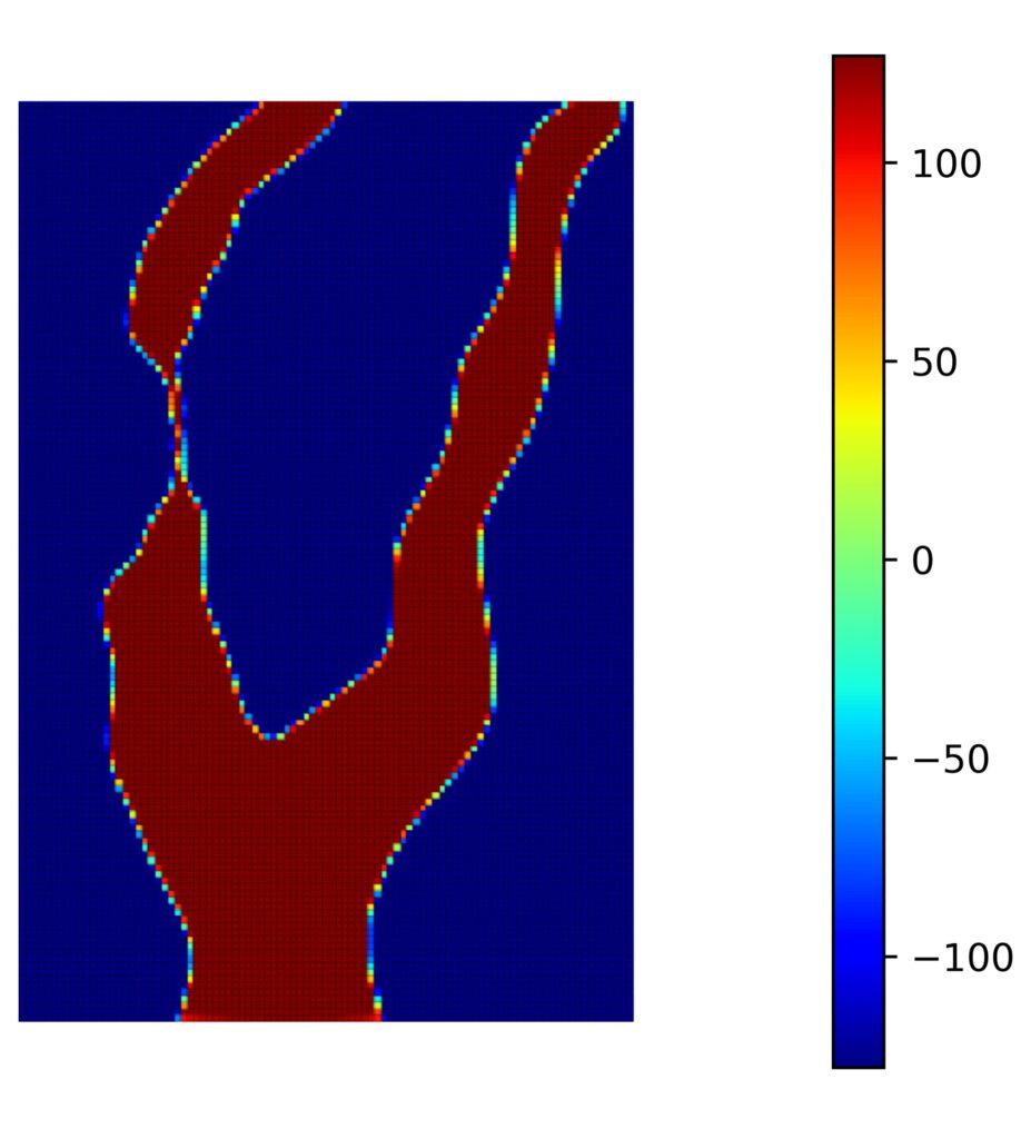
Fig. 1: Voxel data from a gray scale image.
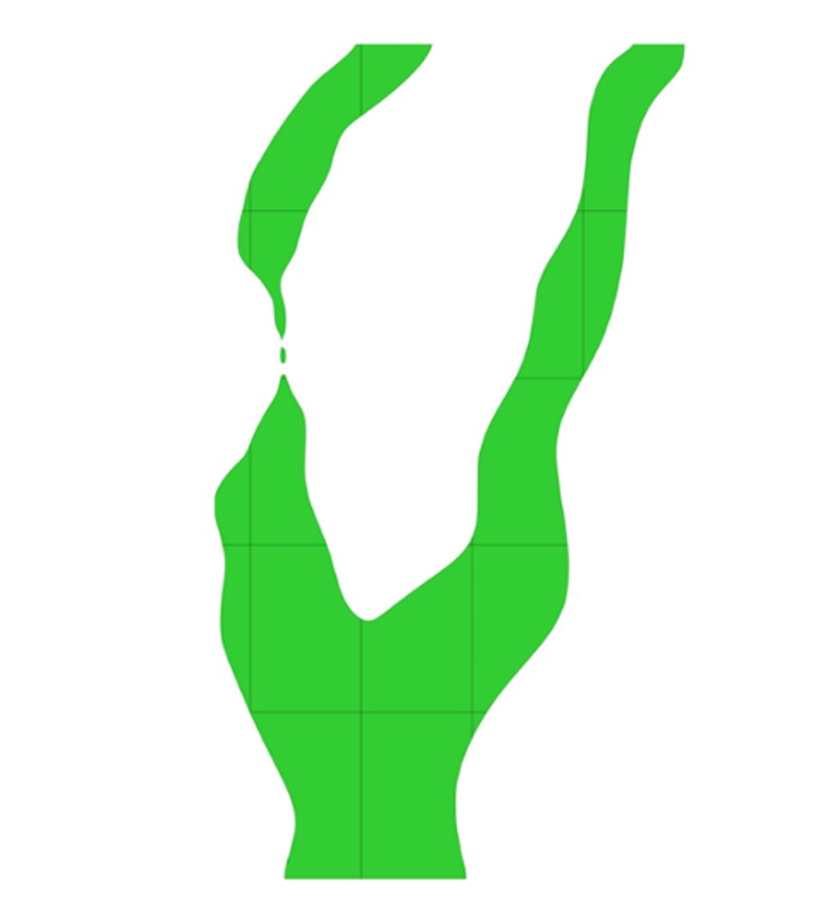
Fig. 2: Reconstructed geometry based on B-spline approximation.
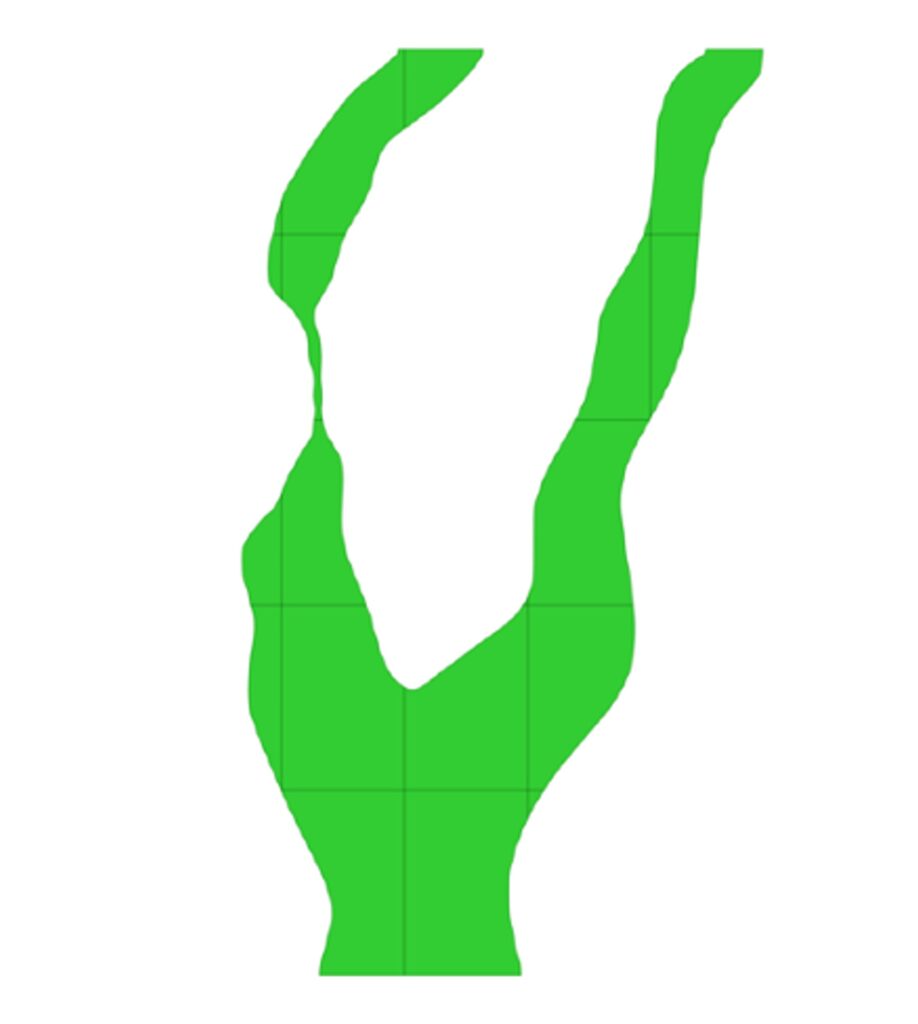
Fig. 3: Reconstructed geometry based on advanced image-segmentation.
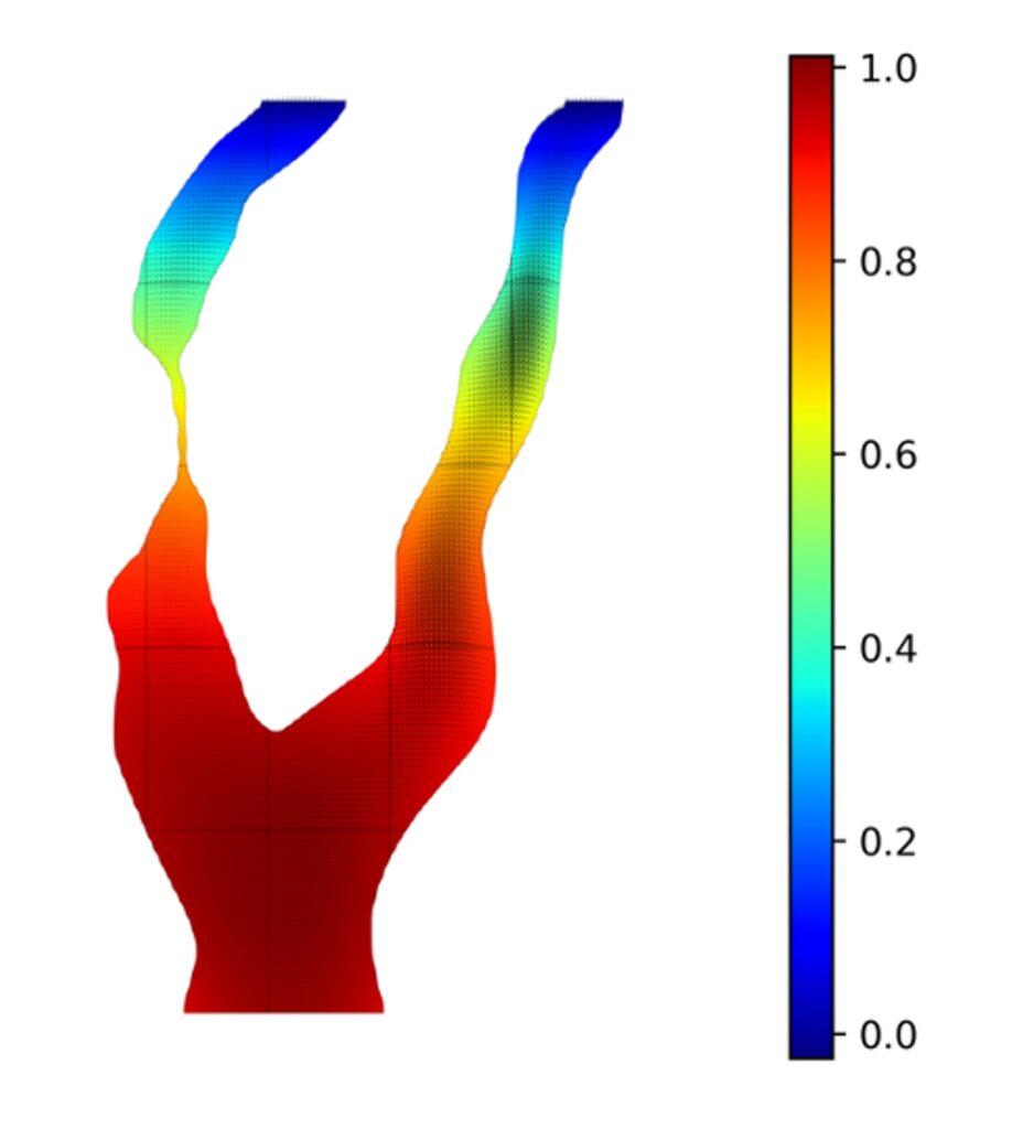
Fig. 4: Stokes flow in a stenotic carotid: Pressure as a colormap and velocity in quivers.
Our CompMech member Sai Chandana Divi, a joint-doctoral candidate between Eindhoven University of Technology (TU/e) and University of Pavia, has been working towards an advanced multi-scale image segmentation algorithm to reconstruct the stenotic carotid (see Fig. 3) under the supervision of dr. ir. Clemens Verhoosel, Prof. Harald van Brummelen, Prof. Ferdinando Auricchio and Prof. Alessandro Reali. In combination with a skeleton-stabilized formulation (Hoang et al. 2018), she could perform a scan-based immersed isogeometric analysis of a (prototypical) stenotic carotid (see Fig. 4) with a small number of elements. She will extend the algorithm toward topological preservation in order to achieve an efficient patient-specific immersed isogeometric analysis. For the specifications of the image-segmentation algorithm feel free to contact her.
September 7th, 2020