
Introduction
More and more physicians recognize that most important factor in Aortic Arch Pathology and in stent-graft failure is likely to be the morphology of the arch itself [2], particularly its angulation. That is why the need of define vessel geometry is increasing a lot in these years.
Goal
The aim of this work is to reconstruct anatomical vascular structures such as, aortic arch and branches, from CT scans provided by: Casa di Cura S. Pio X – Fondazione Opera S. Camillo. Furthermore, using the 3D reconstructions of the vessels, calculate vessels geometry and find out some connections in between pathology and extracted measurements.
Materials and Methods
We start from CT Images of the abdominal area. These images have been provided using a contrast media in order to enhance the visualization of the interested area. Subsequently segmentations, 3D reconstructions and rendering of the images have been performed with a collection of libraries named VTK/VMTK [1] (Visualization Toolkit and Vascular Modeling Toolkit respectively). Segmentation of medical images (DICOM), is the process of dividing an image into sets of pixels. It is used to obtain a new image that is more meaningful and easier to analyze. The result of image segmentation can be used to create image 3D reconstructions with the help of interpolation algorithms (such as Marching Cubes). The 3D reconstructions are the areas of interest used to extract the vessels’ centerlines and their branches that are the references needed in order to measure geometry of vessels.
Results
Active contour segmentation via level set methods is an especially elegant segmentation technique that requires the expert to provide an initialization, set control parameters, and terminate the segmentation. Following segmentation pipeline is introduced across six step:
- Step 1. Segmentation and 3D reconstruction
- Step 2. Centerline calculation.
- Step 3. Branch extraction
- Step 4. Vessel Geometry measurements
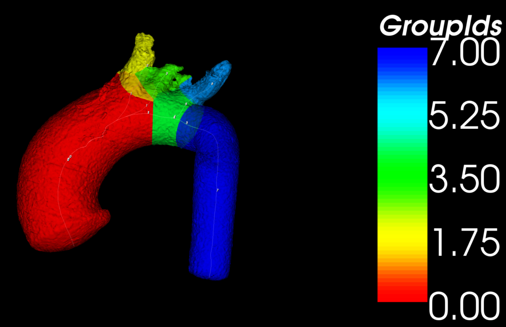
Fig. 1: Aortic Arch 3D reconstruction renderer in opaque with centerline and branch splitting displaying.
References
[1] William J. Schroeder, Kenneth M. Martin, Lisa S. Avila, C. Charles Law. The Visualization Toolkit User’s Guide, Kitware,Inc. 2011
[2] Amir H. Malkawi, Robert J. Hinchliffe, Martin Yates, Peter J. Holt, Ian M. Loftus, and Matt M. Thompson (2010) Morphology of Aortic Arch Pathology: Implications for Endovascular Repair. Journal of Endovascular Therapy: August 2010, Vol. 17, No. 4, pp. 474-479.
Acknowledgements
- Casa di Cura S. Pio X – Fondazione Opera S. Camillo
- Azienda Ospedaliera-Universitaria Senese