
Background
The evaluation of mean clinical parameters before and after percutaneous coronary interventions allows to improve the outcomes of the procedural planning. In particular the mesh generation to image visualization and anatomical features extraction constitute an important aspect in diagnostic field.
Aim
The aim of this study is to provide the surgeon a quantitative and qualitative tool for the measurements of some important clinical features: diameters, lumen area, bifurcation angles and stenosis degree. This study wants to develop a methodological framework to elaborate the images before and after the treatment of coronary lesions.
Materials and Methods
The analyses performed in this study are the following:
- image acquisition;
- image selection;
- image enhancement;
- image segmentation.
Image acquisition. A set of medical images, in DICOM format, are acquired by X-Ray coronary angiography. This technique consists in the radiologic visualisation of the coronary tree through a contrast-dye injections performed from a minimally-invasive catheterization. The images are also correlated to the cardiac cycle by electrocardiographic gating [1]; this approach allows to reduce both the artefacts due to the cardiac motion and to facilitate the choice of the different views at the same cardiac phase, i.e., the end-diastolic phase.
Image selection.Two projection views are selected at the same cardiac phase from the angiographic sequence with a constant CAUD/CRAN angle and varying LAO/RAO angle value. In Figure 1 an example of angiographic images before and after the surgical intervention are shown, which have a pixel resolution of 512×512 and an IPS of 0.2623 mm.
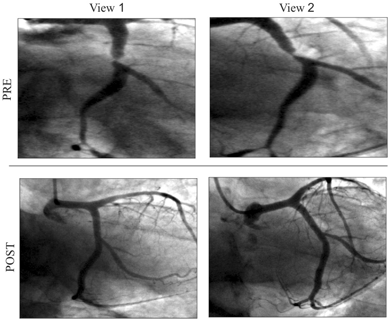
Fig. 1: Image selection of two pair of images about the left coronary artery and its left marginal secondary branch before (PRE) and after (POST) the surgical intervention.
Image enhancement. The image enhancement procedure improves the quality of the image and removes some artefacts presented in the medical images. Such a procedure consists in the manipulation of the original image I(x,y) by the application of specific filters, which are already implemented within the Matlab Image Processing Toolbox. To remove the noise, we apply an adaptive filter, called wiener filter, while for the edges detection in both the views, a Laplacian of Gaussian filtering [2, 3] procedure is computed.
Image segmentation. The procedure consists in the computation of the lumen contours. First, we identify the lumen contours from an user pseudocenter line; the true centerline is calculated from a specific methodology performed in our study. Furthermore, we implement a bi-dimensional analysis of the bifurcation coronary branches and the bifurcation area, separately.
Results
In Figure 2, the results of the image processing is depicted both for the image acquired before the percutaneous coronary intervention and after the procedure to detect the success rate of the surgery, which is important in the clinical practice.
In Table 1 we report the bifurcation angle values of the 3D patient-specific model: A, B and C angle. The A angle, which is the angle between PM and DM, has an influence on the accessibility of the side branch; the B angle, between the PM and SB, is related to the technical success rate of the stent; the C angle, between the DM and SB, as an impact on the risk of the SB occlusion during main branch stenting. The coronary artery modeling and diameter evaluation before and after surgical procedure are highlighted in Figure 3: from the quantitative measurement of the lumen diameter in both the models, we can note that the patency of the coronary lumen is restored.
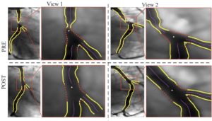
Fig. 2: Results of the image enhancement and segmentation procedure of the two images before (PRE) and after (POST) the surgical intervention. The profile of the lumen contours are changed due the restoration of the lumen patency after the surgical procedure.
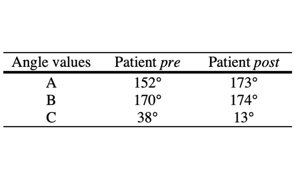
Table 1: Bifurcation angles calculated automatically for the 3D bifurcated coronary model before and after the surgical intervention.
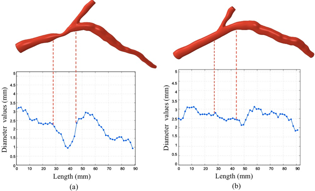
Fig. 3: 3D reconstruction of bifurcation coronary artery before (a) and after (b) the percutaneous coronary intervention. Diameter profile is associated to the respective coronary reconstruction, highlighting the stenosis area in (a) with red line.
Conclusions
In this study we have introduced a simple approach devoted to the image elaboration of X-Ray angiographic images, acquired from the gold standard technique of coronary artery disease before and at the end of the surgical procedure. Providing an accurate bi-dimensional analysis and 3D modeling generation of patient-specific coronary vessel enhance the diagnosis and the treatment, especially in complicated coronary geometries.
References
[1] D. Schafer, J. Borget, V. Rasche, and M. Grass. Motion-compensated and gated cone beam filtered back-projection for 3d rotational x-ray angiography. IEEE Transaction Medical Imaging., 25(7):898–906, 2006.
[2] R.C. Gonzales and R.E. Woods. Digital Image Processing. MA Addison Wesley Publishing Company Reading., 2002.
[3] M. Sonka, V. Hlavac, and R. Boyle. Image Processing Analysis and Machine Vision. Pacific Grove, CA: PWS Publishing., 1999.