
Introduction
Reconstruction methods from DICOM Images represent an important process in clinical medical imaging in order to get a patient-specific analysis of vascular anatomy and a detailed geometry of vascular biomechanics models to improve operative planning. With the recent scienti?c and technological advances, medical imaging is emerging as the most e?ective vehicle of information in health care. The conjugation of 3D anatomical reconstruction from medical images and 3D reconstruction of medical devices is an important tool for surgeon implant simulation and provides pre-operative information to insure the success of medical surgery.
Goal
The research aim is 3D reconstruction of anatomical vascular structures like Carotid Vessel, Aortic Valve Root, Coronary Arteries (RCA, LCA, LAD, CX) from dicom medical images (Computed Tomography Angiography) provided by Policlinico S.Matteo in Pavia. Furthermore, 3D medical devices, like self-expandable stent and balloon-expandable stent for treatment of artery stenosis, are reconstructed from dicom images, which are provided by micro-CT scanner. Tridimensional reconstructions are based on accuracy and precision to ensure high reliability.
Materials and Methods
Methods for quantifying vascular anatomy for patient-speci?c modeling of cardiovascular mechanics include noninvasive imaging techniques such as Computed Tomography Angiography (CTA), Magnetic Resonance Image (RMI), 3D ultrasound (US), and an invasive method combining angiography and intravascular ultrasound (IVUS). Software for image elaboration and analysis is used to visualize and render the surface and volume of patient-specific anatomical structures, such as ITK-Snap, Osirix and 3D Slicer that use a segmentation method. The same software are used to reconstruct medical devices to implant into disease vascular regions of the patient. Image segmentation provides a wide range of biomedical research problems: heart morphology, cancer detection, treatment planning, robot-guided surgery, etc. The spectrum of segmentation techniques available to the clinical researcher is broad, ranging from manual slice-by-slice outlining to fully automatic or semi-automatic techniques that incorporate prior knowledge about the shape and intensity of the structures of interest. Image segmentation is de?ned as the partitioning of an image into non overlapping, connected regions that are homogenous with respect to some characteristic such as intensity or texture. At the end of the segmentation process every pixel in the image is part of a distinct region or segment. However, in the segmentation of electron density maps, it is often more appropriate to remove the constraint that all regions need to be connected. Due to the nature of the data, density below a certain threshold inevitably is noise.
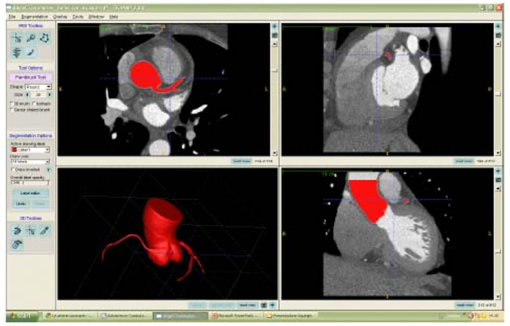
Fig. 1: Itk-Snape interface to image analysis and elaboration. Segmented region of an aortic root with coronary artery (LAD, RCA, LCA) and 3D reconstruction are shown with red color from angio-TC images in dicom format.
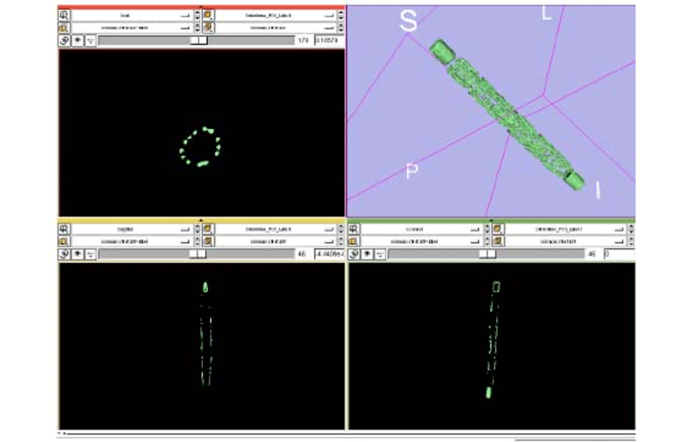
Fig. 2: 3D Slicer interface to image analysis and elaboration. Segmented region of a metallic coronary stent and 3D reconstruction from micro-CT scan in dicom format.
Results
Active contour segmentation via level set methods is an especially elegant segmentation technique that requires the expert to provide an initialization, set control parameters, and terminate the segmentation. Following segmentation pipeline is introduced across six step:
- Step 1. Adjustment contrast of the CT Image
- Step 2. Select the Region of Interest using the Snake Interaction Mode
- Step 3. Construct a Region Competition Feature Image
- Step 4. Initialize the Snake with Bubbles
- Step 5. Update Mesh
- Step 6. Smooth Filter
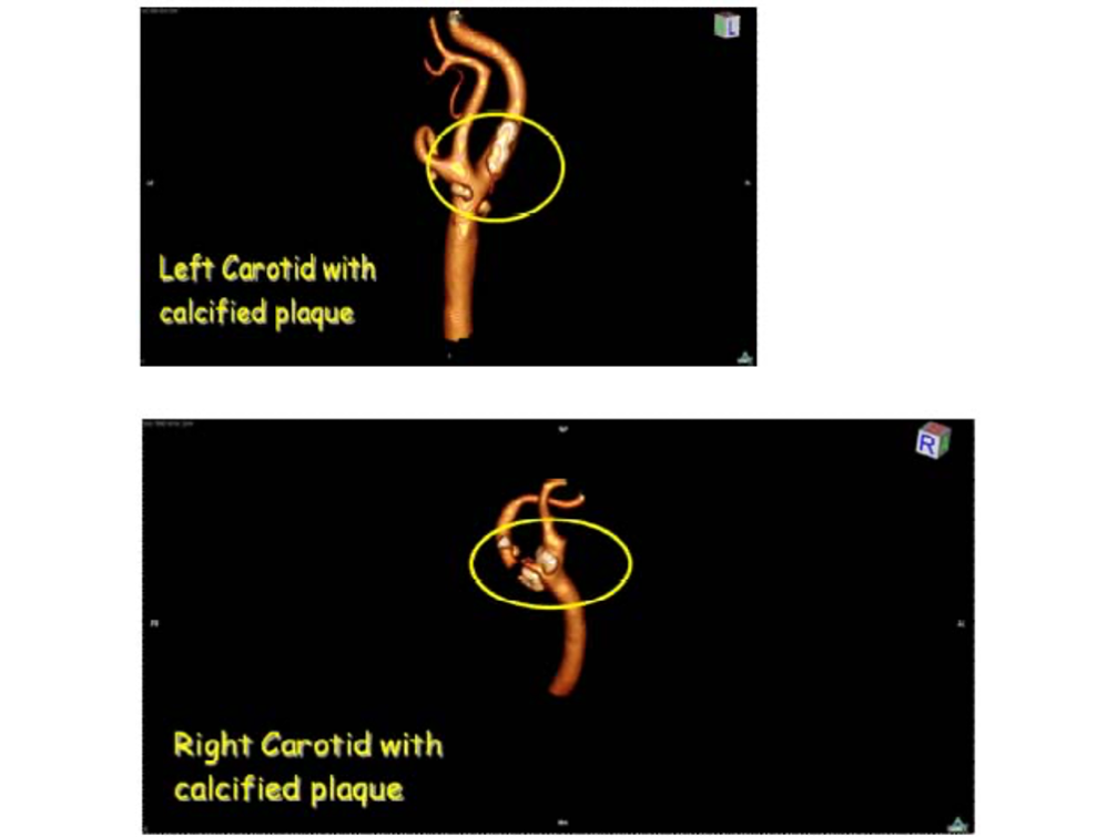
Fig. 6: Left and Right Carotid with calcified plaques near vessel biforcation.

Fig. 7: 3D Stent Device Reconstruction after segmentation pipeline.
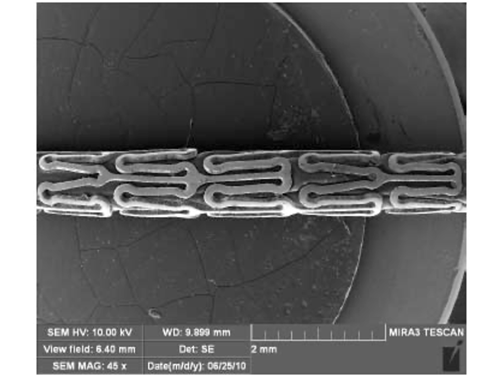
Fig. 8: Stent geometry image from Scanning Electron Microscopy (SEM).

Fig. 3: Modeling cardiovascular anatomy from patient-specific images: aortic root and coronary arteries.

Fig. 4: On the left figure: Identification of principal coronaries: Left Main or Left Coronary Artery (LCA), Left Artery Descending (LAD), Right Coronary Artery (RCA) and Circumflex artery (CX). On the right figure: Stent device into RCA and calcified plaques into LAD and CX.
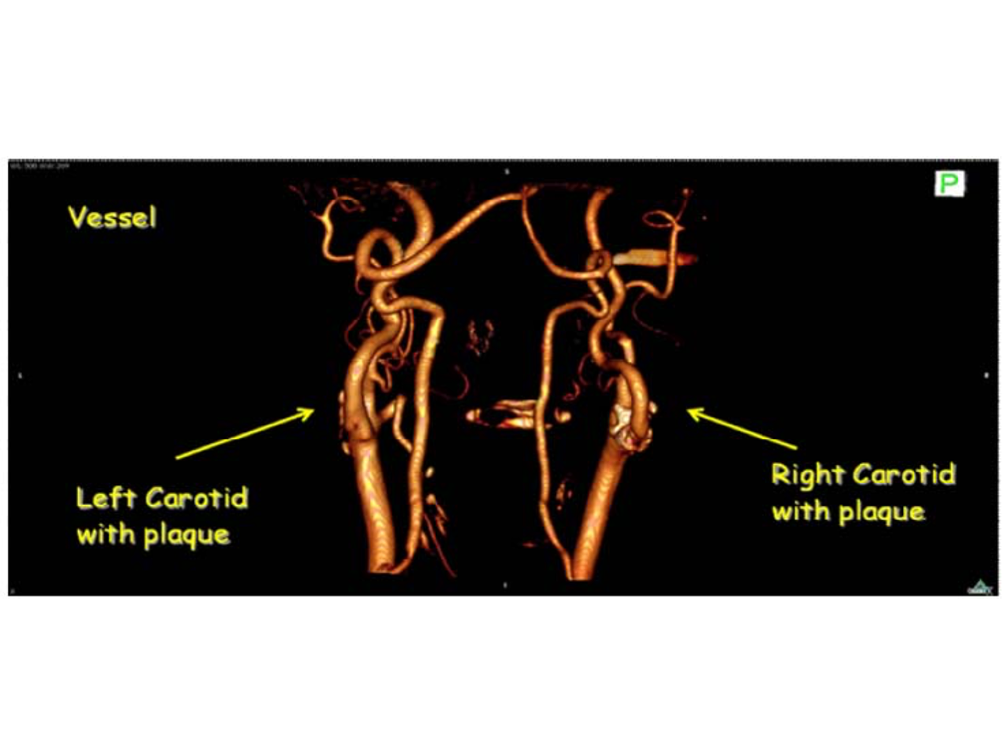
Fig. 5: Reconstruction of vessel cranial tree.
Acknowledgements
- IRCCS San Matteo: Istituto di Radiologia, Dott. R. Dore and Dott. A. Vercelli
- Laboratorio di Farmacologia Molecolare dell’Istituto Mario Negri, Dott.ssa S. Previdi and Dott. M. Broggini
- Laboratorio Arvedi dell’Università degli Studi di Pavia, Prof. M.P. Riccardi and Dott.ssa E. Basso