
Background
Coronary artery disease is the main cause of ischemic heart disease and death in developed countries. Particularly, the treatment of coronary lesions at the bifurcation is a current challenge interventional cardiology. Providing a virtual coronary model and information about stenting implant can represent an important support for the clinical practice, especially for the procedure planning [1].
Aim
The aim of this study is to implement a method for 3D reconstruction of bifurcation coronary arteries to provide a computer-based tool, having a simple graphical user interface (GUI), devoted to the 3D visualization of the coronary vascular tree and to the calculation of its several geometrical features from two single-plane angiography images [2,3,4]. This elaboration can be further extended for the generation of 3D computational mesh of the vessel wall to be used within structural Finite Element Analysis (FEA).
Materials and Methods
In this study we define a methodological framework, resembling the work- flow depicted in Fig. 1.
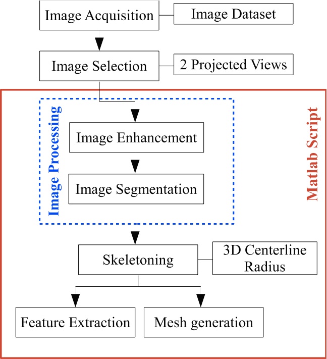
Fig. 1: Schematic work-flow of the 3D reconstruction procedure.
In the following, we describe briefly each step of the workflow, which is implemented in Matlab (The MathWorks Inc., Natick, MA, USA).
- Image enhancement is performed to improve the image quality, removing the noise and enhancing the contrast of the original image [5], as shown in Fig. 2.
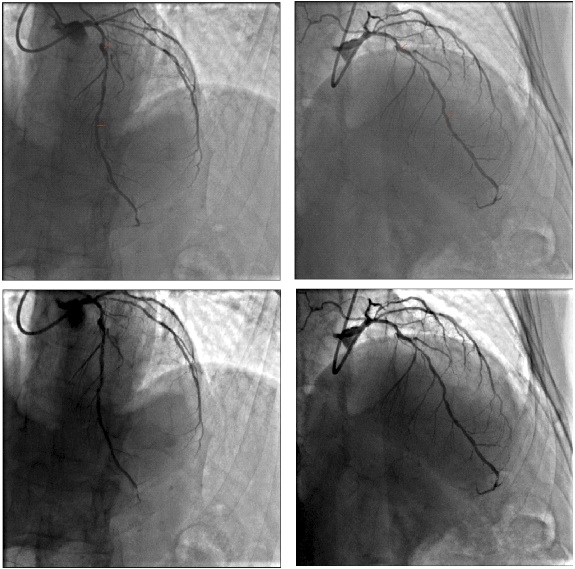
Fig. 2: Image processing from the X-Ray angiographic technique. Up: on the left the first projected view and on the right the second one of the left bifurcation coronary artery. Bottom: the respective views after the image enhancement.
- After the edges detection, an user-defined pseudocenterline is tracked for both views. Segmentation procedure computes the contour points, the centerline, the point of bifurcation and the carinal point. Automatically, the algorithm discerns the three segments of the bifurcation coronary artery, i.e., secondary branch (SB), proximal main (PM) and distal main (DM), as shown in Fig. 3.

Fig. 3: Automatic procedure computes the bifurcation coronary features: PM and DM contours (yellow), SB contour (green), centerline of the three branches (magenta), POB (white) and carina point (cyan) in the first view and second view.
- 2D image analysis is performed to discern the bifurcation region (ROB) and three segment parts of the bifurcation coronary artery to optimize the 3D reconstruction of coronary model. Automatic length estimation of the side branch allows to compute the side branch overlapping on the main vessel. For a correct 3D reconstruction, the procedure overcomes the overlapping problem with the creation of a 2D virtual contours, automatically. In Figure 4, we shown the ultimate contouring procedure of the bifurcated artery [6].

Fig. 4: Image processing: the detected contours follow the lumen edges both in the ROB and the surrounding parts of the lumen.
- 3D reconstruction approach is performed from the information of two single-plane angiographic images, taken from different angle values [7]. 3D centerline and lumen cross-sections are computed from the back-projections of the views. The algorithm computes the vessel wall from the cross-sections enlargement, imposing a variable thickness values along the vessel length. Fig. 5 shown the mesh computation from 2D analysis of the image.
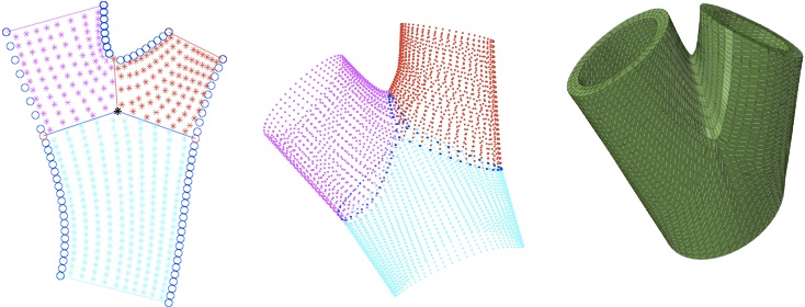
Fig. 5: 3D ROB generation: (a) ROB segmentation and three-different area identification; (b) cross-sections and peanut-like section reconstruction from the extreme sections identification; (c) computational mesh generation.
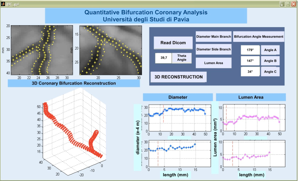
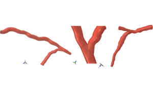
A Graphical User Interface, shown in Figure 6, provides both the morphology and morphometry of the bifurcation coronary arteries, computing the reference diameter, the lumen area and the bifurcation angle [8, 9]. Moreover, we set up a semiautomatic algorithm to generate hexahedral volume mesh for patient-specific stenting simulation, as depicted in Figure 7 .
Conclusions
The present study provides a tool for 2D image processing and 3D model reconstruction from two single-plane angiographic views, supported by a GUI tool. Such a tool, combined with realistic computer-based simulations of coronary stenting may anticipate the surgery outcomes and facilitate the procedure planning.
References
[1] G. A. Sgueglia, D. Todaro, and E. Pucci. Complexity and simplicity in percutaneous bifurcation interventions. EuroIntervention., 6(5):664–5, 2010.
[2] J. Messenger, J. Chen, D. Carroll J.E.B. Burchenal, K. Kioussopoulos, and B. Groves. 3d coronary reconstruction from routine single-plane coronary angiograms: Clinical validation and quantitative analysis of the right coronary artery in 100 patients. The International Journal of Cardiac Imaging, 16:413–427, 2000.
[3] Qin Li, H. Shui, X. Hu, Y. Liu, and Y. Wang. How to reconstruct 3d coronary arterial tree from two arbitrary views. In 3rd International Conference on Bioinformatics and Biomedical Engineering, 2009.
[4] Uwe Jandt, Dirk Schaper, Michael Grass, and Volker Rascher. Automatic generation of 3d coronary artery centerline using rotational x-ray angiography. Medical Image Analysis, 13:846–858, 2009.
[5] R.C. Gonzales and R.E. Woods. Digital Image Processing. MA Addison Wesley Publishing Company Reading., 2002.
[6] Y. Onuma, C. Girasis, J.P. Aben, G. Sarno, N. Piazza, C. Lokkerbol, M.A. Morel, and P.W. Serruys. A novel dedicated quantitative coronary analysis methodology for bifurcation lesions. Technical report, EuroIntervention., 2011.
[7] S. J. Chen and J. D. Carroll. Coronary angiography. Practical Signal and Image Processing in Clinical Cardiology, Springer-Verlag London Limited, Goldberger J.J. and Ng J., Chapter 13, pages 157–185, 2010.
[8] D. Dvir, H. Marom, and A. Assali. Bifurcation lesions in the coronary arteries: Early experience with a novel 3 dimensional imaging and quantitative analysis before and after stenting. EuroIntervention., 3:95–99, 2007.
[9] T. Wischgoll, J. S. Choy, and G. S. Kassab. Extraction of morphometry and branching angle of porcine coronary arterial tree from ct images. American Journal Physiology – Heart and Circulatory Physiology, 297:H1949–H1955, 2009.