
Introduction
The aortic root is a complex anatomo-functional unit that is constituted of the ventriculo-aortic junction, the aortic leaflets, the interleaflet triangles, the Valsalva sinuses with the coronary ostia, the sinotubular junction, and the ascending aorta. The comprehension of the morpho-functional basis of aortic root anatomy and pathology, as well as the possibility of distinguishing the contribution of each single component of the aortic root in either aortic valve dysfunction or in aortic root dilatation, is a major clinical need both for anatomo-functional diagnosis and monitoring of aortic root pathology. 3D reconstruction of MDCT and MR imaging of the aortic root provides morphologic information that contribute to characterize the macro-anatomy of each component of this complex unit. Both MDCT scan and MR have major advantages and limitations: the former is fast but requires high radiation doses and administration of contrast. The latter cannot be performed in a high number of patients due to claustrophobia or permanent devices, etc. Nonetheless, the information that can be obtained from 3D reconstruction can be of major clinical relevance especially for structures that cannot be easily characterized in planar sections. 2D echocardiography is the routine tool for imaging the aortic root, offering the advantages of easy repeatability and no contrast administration. However, morphological information does not provide the contribution on anatomic details as those obtained with 3D reconstruction of MDCT and MR. A major step in progressing though a 2D echo-based 3D reconstruction of the aortic root would open the possibility of evaluating details of aortic root anatomy and monitoring the modifications of both morphology and dimensions.
Goal
The goal is to generate a parametrical rapid off-line 3D morphological representation of the aortic root implemented within a commercial CAD software based on routine 2D transthoracic echo measurements. The proposed procedure should be applicable to both physiologic and pathologic aortic roots and easily extended to bicuspid aortic valves.
Methods
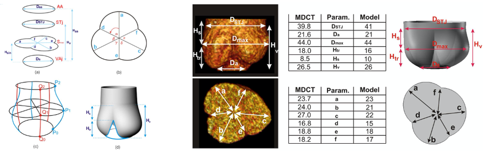
After collecting several patient-specific images of 2D Echo for model generation and of MDCT for model validation, we developed a script within a commercial CAD software which, given few routinely measured dimensions of the aortic valve, is able to automatically generate a 3D asymmetric model of the patient’s valve. The obtained model is validated by comparing it with 3D angio-MDCT scan reconstructions [P1]
Group publications
- [P1] S. Morganti, A. Valentini, V. Favalli, A. Serio, F.I. Gambarin, L. Mazzocchi, D. Vella, M. Massetti, E. Arbustini and F. Auricchio. Aortic root 3D parametric morphological model from 2D-echo images. Submitted to Compuetrs in Biology and Medicine.