
Stroke caused by an embolism accounts for about a third of all stroke cases [1]. Understanding the source and cause of the embolism is critical for diagnosis and long-term treatment of such stroke cases.
The complex nature of the transport of an embolus within large arteries is a primary hindrance to a clear understanding of embolic stroke aetiology. Recent advances in medical image-based computational hemodynamics modelling have rendered increasing utility to such techniques as a probe into the complex flow and transport phenomena in arteries. In this study, CompMech group, in collaboration with the Karolinska Institutet of Stockholm in Sweden, uses computational model to investigate the motion of emboli across a patient-specific ophthalmic artery.
Patient computed tomography image is used to construct a 3D model of the carotid bifurcation. The domain of interest included the common carotid artery (CCA), the proximal part of the external carotid artery (ECA), the ophthalmic artery (OPH) and the internal carotid artery (ICA) before entering the Circle of Willis. The model was discretized into a nominally isotropic tetrahedral finite-element mesh. Subsequent local refinement was performed to enhance mesh resolution in the ophthalmic artery. The overall workflow starting from image data to embolus transport model has been illustrated in Fig. 2.
Blood flow velocities and embolic particle trajectories are resolved using a coupled Euler–Lagrange approach. The embolus trajectory is modelled using Lagrangian particle equations accounting for embolus interaction with blood as well as vessel wall. ANSYS CFD 19.2 is used in the simulations.
The results show that the tendency of large particles to preferentially enter the wider bifurcation branch is augmented under pulsatile flow conditions and the smaller particles are more prone to exit through smaller branches with higher probability to end up in the ophthalmic circulation. Conversely, bigger particles are less likely to follow the fluid inertial forces and thus the flow split.
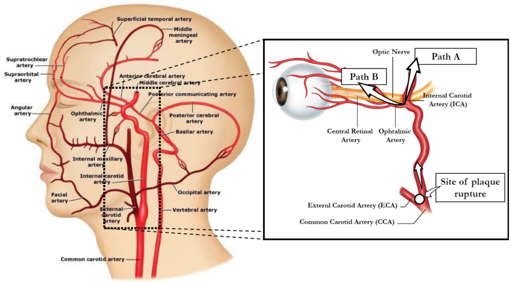
Fig. 1: Path of Carotid plaque. Transient/permanent ischemia (TIA) or minor stroke (MSI) if cerebral circulation is embolized – path A. Amaurosis fugax (AFX) if emboli deposit in the ophthalmic circulation – path B.
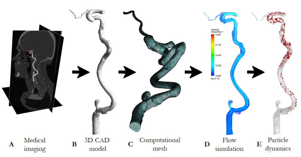
Fig. 2: Schematic overview of the overall computational framework.
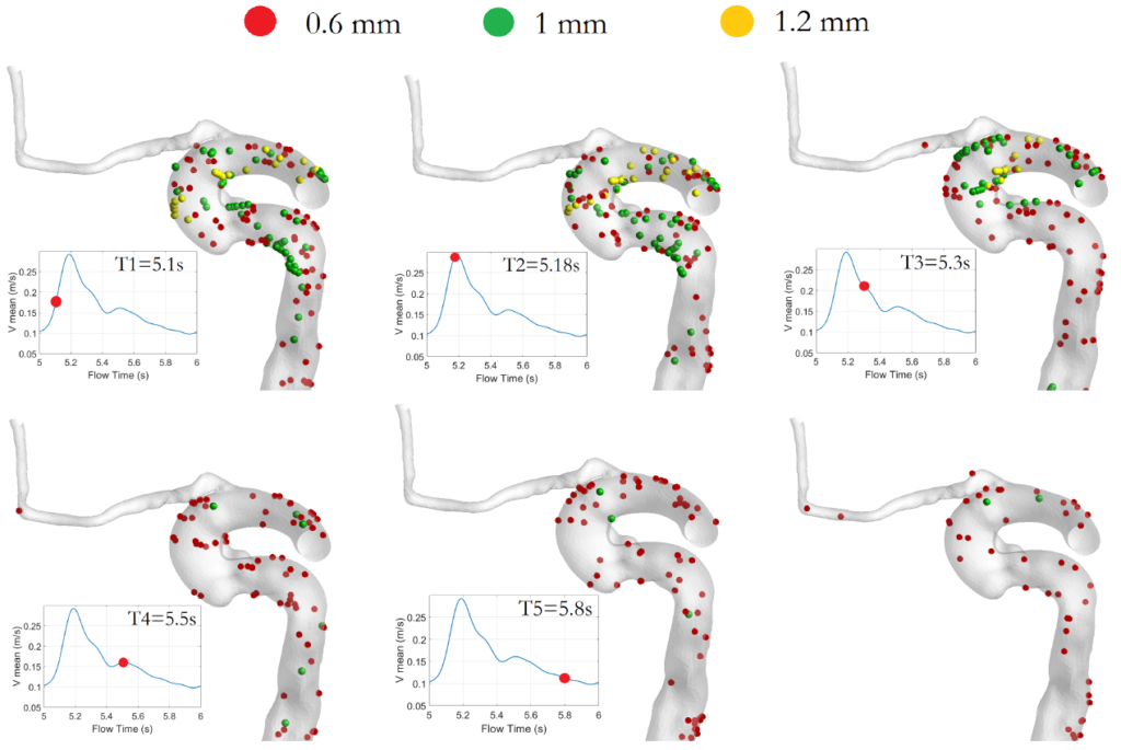
Fig. 3: Snapshots depicting the particle’s location at successive instants during the cardiac cycle. Snapshots (from left to right) are taken at 5.1 s (1), 5.18 s (2), 5.3 s (3), 5.5 s (4), 5.8 s (5), and 7.3 s (6) from the start of the fifth cardiac cycle. The particles are coloured by their diameter.
References
[1] Arboix, A., Alió, J.: Cardioembolic stroke: Clinical features, specific cardiac disorders and prognosis. Curr. Cardiol. Rev. 6(3): 150–161 (2010)
April 2nd, 2020