
Clinical Case Studies
One of the main new frontiers of medicine is to provide patient with a therapy tailored on his specific anatomical and physiological characteristics. On the other hand, the introduction of the 3D printing technology and its wide spread of the last years allow to have in your hands an exact replication of a virtual model.
In this context, image analysis and 3D reconstruction techniques allow to move from the patient CT or MRI to a 3D virtual model of the target anatomy, while the 3D printing allow to move from a virtual to a real anatomical model. The combination of this two skills will pave the way to a complete patient specific pre-operatory planning.
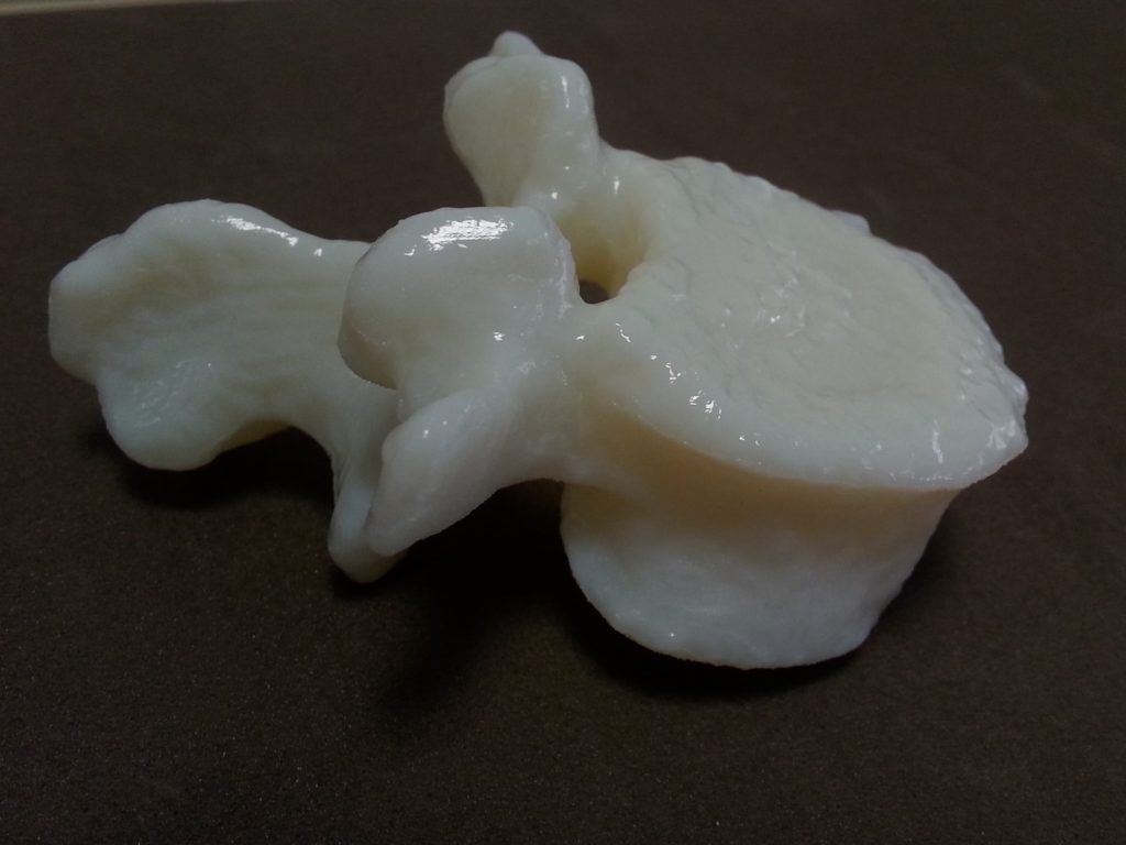
Case-Studies: spinal cord bone
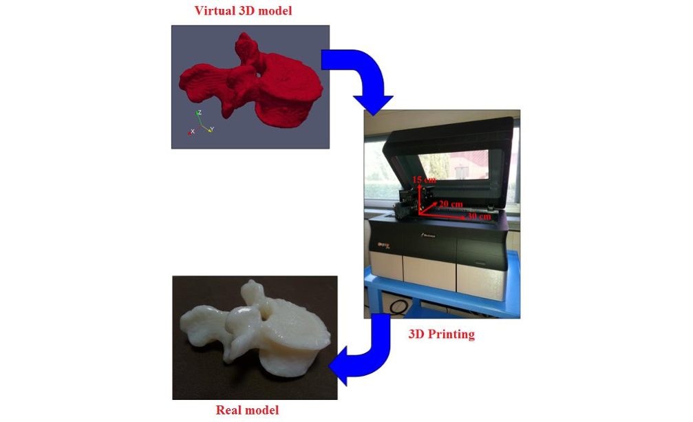
Here we present a simple case study, to explain the prototyping process of a patient-specific anatomy.
We start from volumetric dataset, like CT or MRI and we extract the 3D model using segmentation techniques.
We export the 3D virtual model in .STL format. Then we are ready for the rapid-prototyping with the 3D printer.
In this case the whole process requires 7 hours to move from the patient CT dataset to the final prototyped object.
Obviously, the timing may vary depending on the complexity of the structure to be reconstructed and of the dimension of the final object.

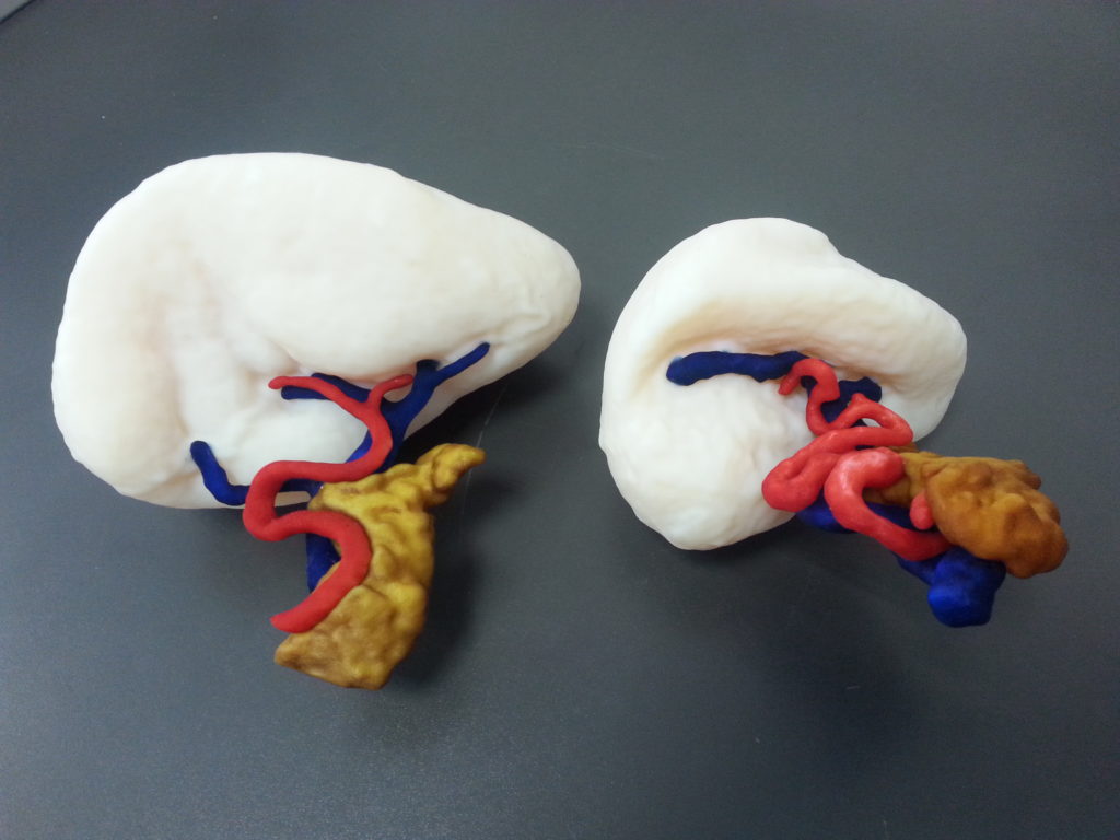
Spleen model: planning of surgical resection.
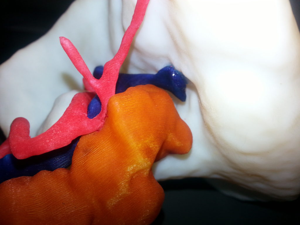
Spleen model, splenomegaly case: planning of surgical resection. Notice the difference between the splenomegaly case and normal case.

Healthy kidney model: planning of surgical extraction from living donor.

Kidney model with a tumor: planning of tumor resection (dark blue).


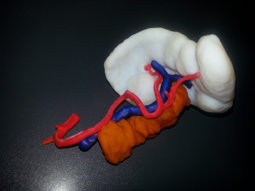
Pancreas model with a tumor at the tail: planning of tumor resection (black).
