
In-vitro and ex-vivo experiments are used as the first step for the validation of ardiovascular medical devices (e.g., stent-graft) [1].
Aiming at the investigation of cardiovascular mechanics in both physiological and pathological conditions, Prof. Michele Conti and Eng. Daniele Bianchi of our group are supporting the reboot of Beta Lab (https://compmech.unipv.it/laboratories/beta-lab/) experimental activities in collaboration with Hospital Policlinico of Milano (https://www.policlinico.mi.it/).
In particular the research activities are focused on the evaluation of the effects of thoracic endovascular aortic repair (TEVAR) on aortic biomechanics. Several specimens of porcine aorta (segment between the aortic root and the renal arteries) was connected to the developed pulsatile mock loop system [2]: intraluminal pressure was recorded in order to measure pulse wave velocity before and after the stent graft deployment. The experimental findings give a clinical insight for both computational modeling and therapeutical approaches.
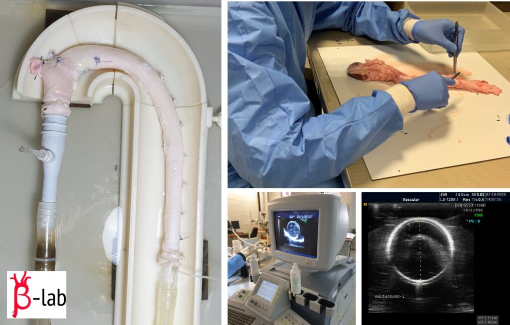
Fig. 1: An overview of the experimental activities regarding the evaluation of the effects of thoracic endovascular aortic repair (TEVAR) on aortic biomechanics .
References
[1] H.W.L. De Beaufort, M. Conti, A.V. Kamman, F.J.H. Nauta, E. Lanzarone, F.L. Moll, J.A. Van Herwaarden, F. Auricchio, S. Trimarchi. “Stent-Graft Deployment Increases Aortic Stiffness in an Ex Vivo Porcine Model”, Annals of Vascular Surgery (2017) – DOI: 10.1016/j.avsg.2017.04.024
[2] E. Lanzarone, R. Vismara, G.B. Fiore. “A new pulsatile volumetric device with biomorphic valves for the in vitro study of the cardiovascular system”, Artificial organs (2009)
April 1st, 2021