
Aortic lumen segmentation is a critical step to assess the aortic anatomy, identify pathology, and perform preoperative planning in vascular surgery. During preoperative planning, the aortic lumen is extracted from Computed Tomography Angiography (CTA) in order to measure aortic length and diameters thus enabling the endograft sizing and/or to perform finite element analysis. Currently, aorta segmentation is performed using manual or semi-automatic tools that are time consuming and provide results that are operator dependent and difficult to reproduce. In recent years, deep learning has shown excellent performances in medical image analysis, exploiting Convolutional Neural Networks (CNN) for segmentation, classification, and detection tasks.
Carried out in cooperation with Camelot Biomedical Systems S.r.l (Genoa) and the Department of Experimental Medicine (DIMES), University of Genoa, the project has focused on deep learning approaches to locate and segment the whole aortic lumen from thoracic aorta to the iliac arteries accounting for spatial consistency.
In particular:
- A first 2D CNN is used to coarsely segment and locate the aortic lumen from the whole CTA scans at a lower resolution
- Three orthogonal 2D CNN are used to segment the region of interest (ROI) identified in the previous step at a higher resolution on the axial, sagittal, coronal planes respectively
- The results provided by the orthogonal networks are combined together to provide a final segmentation that is spatially coherent, overcoming the limitations of single-view CNN
The obtained results show that the proposed approach can accurately localize and segment the aortic lumen in subjects with aneurysm. Moreover, the integration of CNNs trained on axial, sagittal, and coronal views provides better results than single view segmentation and enforces spatial coherence.
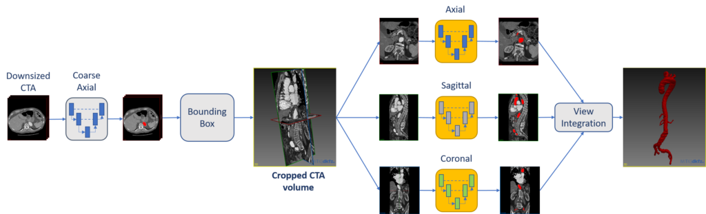
Fig. 1: The first step of automatic segmentation involves the coarse localization of the aortic lumen on low-resolution axial images. In the next step, models trained on axial, sagittal and coronal planes are exploited to perform single-view segmentation from the ROI identified in the previous phase. Finally, the segmentation is obtained by integrating the three orthogonal segmentations.

Fig. 2: Ground truth (in green) and predicted segmentation (in red) are overlaid on the CTA. The automatic segmentation is accurate.

Fig. 3: The ground truth 3D model and the model generated by the automatic segmentation are represented in green and red, respectively.
Since lumen segmentation is the first step for morphology analysis and stent-graft sizing, this automatic pipeline can significantly decrease the workload for surgeons in the planning stage. Moreover, the ability to segment the aortic lumen from thoracic to iliac areas enables different types of geometric analysis (e.g., iliac tortuosity estimation, thoracic aortic arch analysis, etc.)
April 16th, 2020